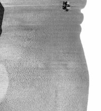
Liz Orton uses medical imaging and clinical representation to focus on the medical gaze and its power relations. Through collaborative portraits, co-opted algorithms, and appropriated and cropped images from medical textbooks, she seeks to reclaim what makes us human from the hard facts and figures of medical imaging and the ways of seeing that accompany them.
It’s work that humanises images that are seen as evidential, scientific and objective yet come with a history that is tied to ideas of institutional power, surveillance and male dominated hierarchies. In that sense, her work is not just about the medical gaze, it’s about the history of photography and the ways in which it is used against people.
It’s also work that has a personal edge, its roots sunk in Orton’s experience of a ‘non-invasive’ MRI scan her 13-month-old daughter had to undergo at Great Ormond Street Hospital.
“They wanted to understand function through an imaging procedure so they injected her with a dye and they told me in a very matter-of-fact way that she would have to be motionless for 15-20 minutes,” says Orton in a Skype interview from her home in London. “This was a 13-month-old baby, remember, to be kept still for 15 minutes.”
“I tried feeding her to keep her still and that worked for about 3 minutes, but in the end they had to strap her to the bed with sandbags and these massive things of Velcro,” says Orton. “What does that mean for their understanding of what the body is when you say that is not invasive. What they mean is there is no cutting, no physical cutting. By suggesting that procedure is non-invasive, it says in their definition of the body, that the body is a purely physical object, continuing this whole separation of the body and the mind, and the idea that the body and psycho-emotional affects are unconnected.”
Orton’s interest in the medical gaze developed through participatory photography workshops she ran in community settings where the illness of the participants formed a common thread. This work led her to running photography courses with medical students, using photography to help them see the body in a way that questioned the usual medical view of the body and the idea of the heroic doctor as all-seeing conqueror of disease.
“The histories of photography and medicine are so entangled so I got to think about it practically but also to examine theoretical ideas about objectivity, and the construction of knowledge. I wanted to get them to think about the way they are taught about the body, the way they think about the body, and how the photographer-subject relationship in a practical and theoretical sense is a metaphor for medical ways of seeing. The ethics of looking and consent are a huge part of that.”
Orton’s interest in the body and the way it is represented in medicine has crystallised in her latest projects, work that resulted from a grant provided by the Wellcome Trust. Using images from radiography guides and patient scans, Orton has created a body of work that seeks to reclaim the subject from the medical gaze.
“One of the things I did early on was refuse the term the medical body,” she says. “It implies that we have different bodies; cultural bodies, social bodies, medical bodies. And that’s part of the problem. It’s the same body.”
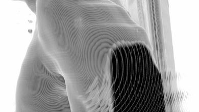
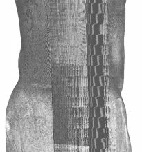
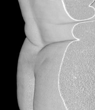
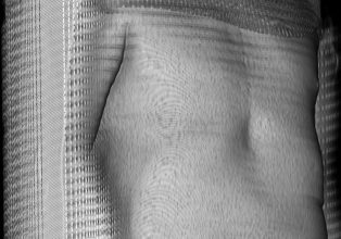
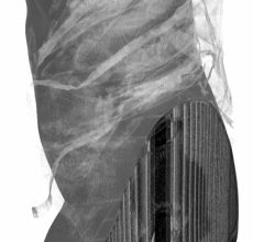
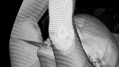
For her ‘Digital Insides’ project, Orton spent time in imaging departments, co-opting visualisation algorithms and using patient data to make images with a more human aspect. For this project, Orton would go down into the bowels of the hospital (x-rays use a lot of protective lead so radiography departments are often in hospital basements) to observe the technicians capturing the data that would determine the patients’ clinical condition. Later, Orton got the patients’ permission to use that raw data to rebuild the patient from a clinical subject to something more human.
Reclaiming the medical gaze
“My work is about reclaiming the medical gaze. So I learned how to use radiological software, partly by watching radiologists in these cell-like rooms. I worked with the software to build up three dimensional renderings of people. But where imaging is interested in the inside of a person, I wanted to draw a picture of the outside.”
“I wanted to refuse what was inside. So I co-opted the software to subvert the medical gaze and I have these three dimensional layered images, but they have shiny skin, they have little ridges where the data is interpolated. They are a step up from a picture of the interior but I wouldn’t say I like them .”
There’s the idea of the perfect view in landscape painting. There’s also the idea of the perfect view – a kind of data sublime – in computing; one MIT scientist saying that the perfect view for a computer was the inside of the body. “I think I have moved forward from that,” says Orton, “but it’s not very far forward. It’s still using the language of the medical gaze so you have these fractured personalities.” This rebuilding is also connected with the idea of x-rays or MRI scans being a way of seeing. Orton sees this as a metaphor to humanise a process that is closed and has little trace of the human about it.
“When people talk about computer vision, machine vision, it’s a further extension of the visual as a metaphor, because computers are not really seeing; they’re calculating, analysing and interpreting patterns. They are working with code, they’re not looking at anything. It’s a metaphor. Everyone talks about machine vision but I haven’t seen anyone question the idea that it’s about vision. It’s not.”
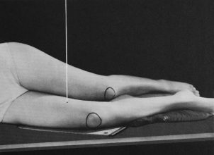
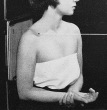
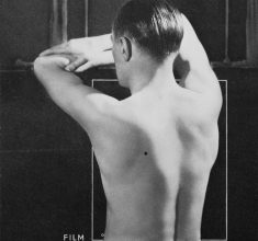
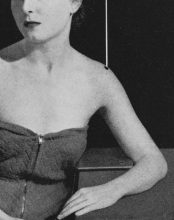
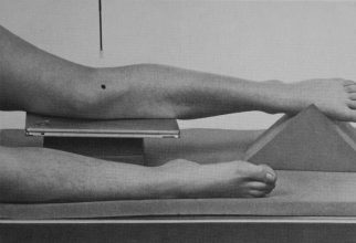
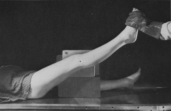
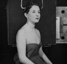
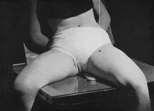
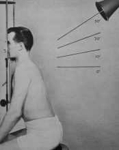
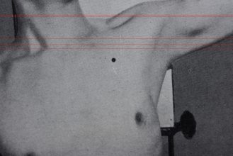
Another side to Orton’s work can be seen her appropriations from the X-ray manual, Clark’s Positioning in Radiography. “Clark’s Positioning for Radiography is a handbook for radiologists on how to position parts of the body for x-rays. The region of interest has to be parallel to the plate, the body is surrendered to the language of the plane, and medicine’s geometry imposed as lines and angles on top of the figure. There are thousands of images, a catalogue of imaged bodies, everyone a diagram or instruction. These images exceed (or refuse to be limited by) their scientific designation. Many of them are charged with such eroticism, comedy or subjection that they tell as much about photography and power as they do about the medical body.”
Using a range of strategies that mirror x-ray framing and angle, she strips away the clinical detail from the image of models in the book, creating images where underlying themes of sexuality, voyeurism sadomasochism and glamour come through. These are hypnotic images where 1950s haircuts feature with dreamy eyed models who look like they’ve stepped out of a Hitchcock movie. There are bubble perms, tops with zippers up the front, and a certain self-consciousness that combines eerily with the lines signifying the track of the X-ray. By pulling them out from their handbook origins, Orton reveals the non-neutrality of the image to the surface, turning the textbook image into a kind of conceptual pin-up. And that is what her work is about, bringing the body to the surface, creating a holistic view of what it is to be human, especially when the tools with which you are being looked at have no humanity to them.
“Photography’s so tied up with ideals,” she says. “I guess one of the things I realised the other day is that maybe through these dialogues I’m doing I’m trying to invert this gaze. I’m trying to release the medical image from these shackles of being determined and defined as an institutional record. The big question is what happens when you pull those images away from those functions and institutional rules and subject it to a different kind of questioning and analysis.”
__________
With thanks to Liz Orton and Colin Pantall for this text
For further viewing: lizorton.co.uk